X-ray, CT uncover novel coronavirus-infected pneumonia
By AuntMinnie.com staff writers
January 27, 2020 — The chest radiographs and CT scans of individuals infected by the novel coronavirus display pneumonia-like patterns that can aid in diagnosis, according to a case report by the Chinese Center for Disease Control and Prevention published online January 24 in the New England Journal of Medicine.
For their investigation, researchers from various governmental and healthcare institutions throughout China analyzed the lung samples of three patients suspected of being infected with the new betacoronavirus (2019-nCoV). The coronavirus has been linked to the Huanan seafood market in Wuhan, and studies into the outbreakare ongoing.
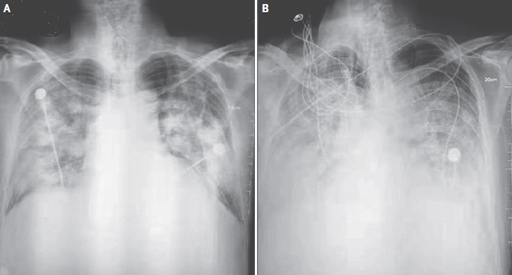
Each of the patients presented with a combination of fever, cough, and chest discomfort. The patients underwent chest x-ray and CT exams that showed pneumonia-like findings, which the researchers referred to as “novel coronavirus-infected pneumonia.” The radiographs of the one patient who died showed an increase in density, profusion, and confluence of bilateral opacities over time, in addition to the accumulation of pleural fluid.

DNA sequencing of bronchoalveolar-lavage samples later confirmed the genome of 2019-nCoV. Electron micrographs of the coronavirus particles showed that they were spherically shaped with some pleomorphism.
The combination of medical imaging techniques, transmission electron microscopy, and whole-genome sequencing allowed for “visualization and detection of [a] new human coronavirus that can possibly elude identification by traditional approaches,” co-first author Na Zhu, PhD, and colleagues wrote. “Further development of accurate and rapid methods to identify unknown respiratory pathogens is still needed.”
