CT links vaping to ARDS in new case studies
By Abraham Kim, AuntMinnie.com staff writer
December 17, 2019 — Radiologists identified vaping-related lung injury patterns on the CT scans of patients with acute respiratory distress syndrome (ARDS) in two recent reports out of New Jersey and Missouri. Early recognition of these imaging patterns could prove vital to timely and appropriate treatment for these patients.
Thousands of EVALI reports have emerged this year alone, and the medical community has identified “pulmonary manifestations of severe disease” from vaping that have resulted in lengthy hospitalizations and dozens of deaths, noted Dr. Nicole Sakla and colleagues from Newark Beth Israel Medical Center.
In their case study, the New Jersey radiologists examined a chest x-ray and CT pulmonary angiogram of a 25-year-old female who presented to the hospital emergency department with shortness of breath, dry cough, and pleuritic chest pain. The medical team diagnosed her with atypical “walking” pneumonia and eventually sent her home with antibiotics after CT and lab tests ruled out pulmonary embolism and various other diseases (Emergency Radiology, December 9, 2019).


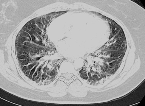
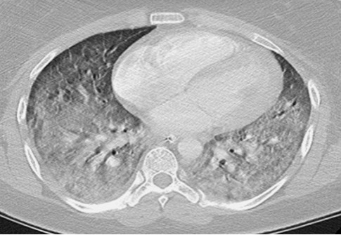
Two days following discharge, the patient returned to the emergency department with worsening symptoms. Chest CT scans revealed signs consistent with diffuse alveolar damage — including extensive opacification of the lung parenchyma with interstitial edema — which is a common mechanism of injury for ARDS. The patient had no previous medical history but reported having used e-cigarettes for about two to three hours per day, three times a week over the past year. Confirming EVALI in the patient convinced the clinicians to send her to another medical center for oxygenation.
Early radiologic recognition of vaping as a risk factor for ARDS is fundamental in emergency cases, Sakla and colleagues noted.
“The consideration of vaping-induced lung disease in a patient with known e-cigarette use should therefore be added to the lexicon of differentials a radiologist considers when evaluating such patients in order to ensure immediate respiratory support and treatment,” they wrote.
A separate team, led by Drs. Raed Qarajeh and Jacqueline Kitchen from the University of Missouri-Kansas City, also uncovered vaping-associated ARDS in a 47-year-old man who had been using e-cigarettes loaded with THC concentrates three times a day (American Journal of Medicine, November 30, 2019).
The patient’s initial chest x-rays showed perihilar and bibasilar heterogeneous opacities, for which he received various antibiotics. His condition continued to worsen, however, and he was transferred to an intensive care unit for supplemental oxygen.
Subsequent chest CT scans revealed that he had multifocal ground-glass and nodular opacities with diffuse interlobular septal thickening, indicative of EVALI. The physicians responded to this finding by giving the patient steroid medication, after which he gradually improved.
Medical professionals should consider EVALI in patients who present with acute hypoxic respiratory failure and other symptoms indicative of ARDS, Qarajeh and Kitchen wrote. As the EVALI epidemic is still in the early stages, there is a need for further investigation into the vaping agents responsible for this type of lung injury, particularly the effect of adding THC to e-cigarette fluids.
