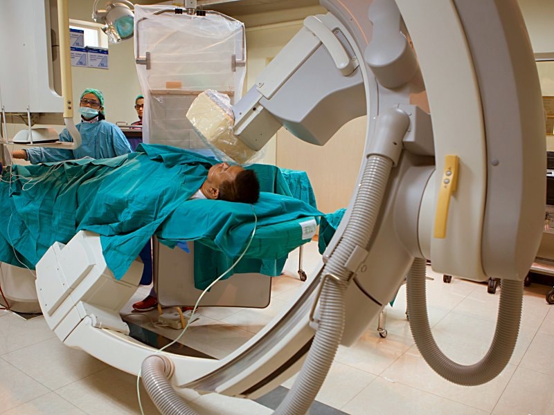
DeFACTO Result: CCTA, Standard Angiography Similarly Prognostic
Pam Harrison
February 24, 2016
RELATED LINKS
 What Strategies Work for Reducing Inappropriate Cardiac Imaging?
 CT-Angiography Beats Perfusion Scan for CAD Diagnosis: Study
DeFACTO Study of Noninvasive Fractional Flow Reserve
My Alerts
Click the topic below to receive emails when new articles are available.
RELATED DRUGS & DISEASES
Percutaneous Coronary Intervention
Clopidogrel Dosing and CYP2C19
Acute Coronary Syndrome
TORRANCE, CA — The insights provided by coronary CT angiography (CCTA) are similar to what is available from quantitative coronary angiography (QCA), suggests a prospective comparative study that gave CCTA the edge because it’s also noninvasive and offers some other advantages over radiographic angiography[1].
The study is described as “the first prospective, head-to-head multicenter comparison” of CCTA and QCA against what was taken as the gold standard, invasive measurement of fractional flow reserve (FFR).
The follow-up analysis of the previously published DeFACTOtrial suggests that “CTA may be used as an alternative to assess luminal stenosis and to serve as gatekeeper to FFR measurements in patients presenting with chest-pain syndromes,” according to the report published February 17, 2016 in JACC: Cardiovascular Imaging.
“Clearly, invasive angiography is going to be the way we are going to fix lesions when people need a stent,” lead author Dr Matthew J Budoff (Los Angeles Biomedical Research Center, Torrance, CA) told heartwire from Medscape. “But when you are talking about diagnosis, especially in a stable patent where there is no urgency, CCTA offers a lot of advantages in that we can see plaque, we can see if there’s been an infarction, we can see other structures like atrial-septal defects or other structures beyond the coronary arteries, including the pulmonary arteries, so we can look for pulmonary embolism at the same time,” he added.
“So CTA provides a big opportunity for emergency-department physicians and cardiologists to get to the true cause of chest pain a lot more quickly than our current approaches. And CT angiography is about 1/20th of the cost of cardiac catheterization, so we can direct our care much better and save a lot of money.”
As the authors note, DeFACTO was designed to evaluate the accuracy of FFR measurements derived from computed tomography (FFR-CT) to diagnose hemodynamically significant CAD as measured by direct-FFR at invasive angiography.
Sending the “B Team”
In an accompanying editorial[2], Dr Armin Arbab-Zadeh (Johns Hopkins University, Baltimore, MD) notes the current results are important “because they demonstrate yet again that conventional angiography is not superior to CT for the diagnosis of coronary artery disease—in this case hemodynamically significant coronary artery disease—when compared with an independent reference standard.”
He adds, “Their results are even more impressive when considering that the authors used only visual lumen assessment for their analysis while conventional angiography employed quantitative evaluation.”
Moreover, the study used only “percent stenosis estimates by CT but did not take advantage of the abundant options for coronary artery disease assessment,” which include assessments of lumen area, area stenosis, lesion length, and plaque burden, Arbab-Zadeh writes.
“Thus, CT sent only its ‘B’ team and still held its own compared with our current gold standard for the diagnosis of coronary artery disease.”
Prospective DeFACTO Comparison
The cohort of 252 patients referred for coronary angiography, mean age 63 years, underwent all imaging procedures for 407 lesions. Lesions of 50% severity or greater were considered to be obstructive.
About half the group, or 54.4%, showed a functionally abnormal lesion by invasive FFR; 37% of all measured lesions in the cohort were found to cause ischemia. QCA and CCTA were similar in their ability to detect lesion-specific ischemia.
Prediction of Lesion-Specific Ischemia
| Measure | CCTA (%) | QCA (%) |
| Diagnostic accuracy | 69 | 71 |
| Sensitivity | 79 | 74 |
| Specificity | 63 | 70 |
| PPV | 55 | 59 |
| NPV | 83 | 82 |
PPV=positive predictive value
NPV=negative predictive value
Area-under-the-curve (AUC) findings assessing overall accuracy of the two tests were also similar, at 0.75 for CCTA and 0.77 for QCA. Per-vessel analysis was also not significantly different than per-patient values.
Given that “CT is capable of delineating atherosclerotic plaque characteristics and estimating total disease burden, which show promise for advanced risk stratification in patients,” Arbab-Zadeh asks, how is it possible that “we still puncture arteries and advance catheters in aortas of more than 1,000,000 patients every year for the diagnosis of coronary artery disease? Simply because we have no data on patient management based on a CT-guided management directly compared with the standard approach of invasive angiography.”
He concludes, “Noninvasive coronary angiography has come a long way and is ready for prime time.” Now, “we have to complete the last and most difficult step, proving its utility in patient management compared with the current standard of conventional angiography.”
Budoff reports serving as a consultant for Heartflow. Disclosures for the coauthors are in the report. Arbab-Zadeh reports no relevant financial relationships.
