Pancreatic Cancer Screening Checklist to Identify High-Risk Patients

Pancreatic Cancer Screening Checklist to Identify High-Risk Patients


Heart CT Scan -First for Chest Pain: The Anatomical Truth Machine

CT-First for Chest Pain: The Anatomical Truth Machine

photo of Jonathon Leipsic
Jonathon A. Leipsic, MD

By 2050, the prevalence of total cardiovascular disease in the United States is projected to grow from 11% to 15%. That growth is already felt by doctors, as nearly 8 million patients annually show up to emergency departments with acute chest pain. When those patients present, we need a quicker, more effective way to test for cardiovascular disease.

Currently, the two tests most often performed are stress testing and cardiac catheterization.

While effective, these tests cost time, money, and effort — sometimes, only to find out they were unnecessary. Too many patients endure slow, costly, invasive pathways before we even confirm that they have coronary artery disease(CAD).

That is why I believe that the first step should be coronary CT angiography(CCTA).

A New Standard of Care

We can do better than relying on stress testing and symptom-based referrals as a first line of defense. Chest pain alone is an imperfect indicator of CAD, and sending patients for catheterizations on the basis of this symptom may end up putting them through unnecessary duress. Right now, we’re seeing 1 million cardiac catheterizations performed annually. We could avoid a fair amount of those if we prioritize a CCTA-first approach.

For over two decades, I’ve been passionate about cardiac CT. This advanced imaging technique uses a lower dose of radiation with imaging conducted within a single heartbeat that scans the entire heart. We receive a real picture of what goes on inside the coronary arteries, quickly revealing what we’re treating. This allows us to proactively identify problems and provide patients with the treatment they need.

Noninvasive and fast, CCTA allows for the confident diagnosis of atherosclerosis and anatomical stenoses, enabling more effective titration of medical therapy and referral to invasive angiography, leading to improved revascularization. It opens a door to the entire picture of a patient’s condition instead of just cracking open a window, helping us diagnose CAD, informing us of any patient risks, and improving medical management.

Why CT?

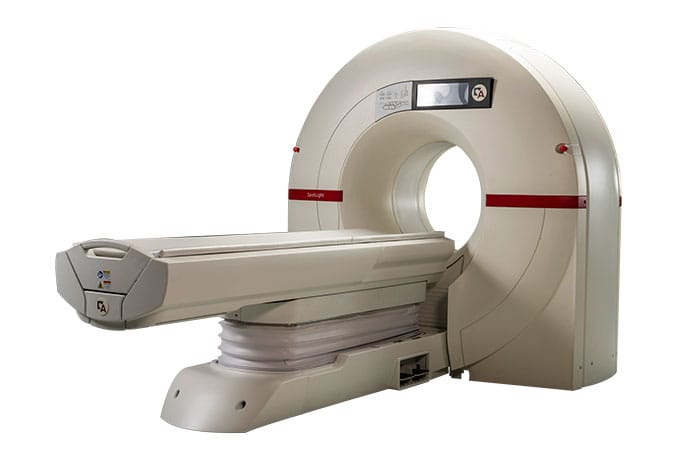
I’ve seen the effectiveness of a CT-first approach firsthand. Back in 2019, we installed a dedicated, small-footprint CT from Arineta at the University of British Columbia. To this day, I’m amazed when I walk into our CT scan room and, instead of a vast unit, see a much smaller piece of equipment on hand to provide the answers.

photo of a CT scanner
Arineta CT Scanner

At the time, I believed that we’d see a rapid adoption of CCTA, but it wasn’t until the 2021 ACC/AHA chest pain guidelines were released that we saw it take off.

Published jointly by a number of professional societies, including the American Heart Association (AHA) and the American College of Cardiology (ACC), the guidelines are used by a wide range of healthcare professionals and organizations, such as the Centers for Medicare & Medicaid Services. Cardiac CT received a Class I, Level A recommendation, validating it as a frontline strategy for diagnosing CAD, supported by evidence from clinical trials.

This top-tier recommendation for cardiac CT as the preferred initial strategy for patients with chest pain and no known CAD is all the more reason to embrace a CT-first approach.

Looking to the Future

We can improve on the current method of sending patients for stress testing or waiting on cath lab appointments; just look at the 50% of patients we’ve seen incompletely revascularized in the ISCHEMIA trial. By implementing a CT-first approach, we can enhance patient outcomes and experiences, while saving them time and money.

The recent position paper  from the Society for Cardiovascular Angiography & Interventions and the Society of Cardiovascular Computed Tomography, which I co-chaired, highlights opportunities to integrate noninvasive physiology, such as fractional flow reserve (FFR-CT) and AI-enabled quantitative atherosclerosis measures into CCTA. We are also seeing growing integration of quantitative plaque assessment in prevention clinics. This broader application through the integration of advanced analytic tools should drive further improvements in clinical outcomes.

I’m confident that cardiac CT will be a standard in cath labs and prevention clinics within the next 10 years.

Jonathon A. Leipsic, MD, is a professor and chair of radiology and a professor of cardiology at the University of British Columbia, and previously served as Canada Research Chair of Cardiac Imaging. Dr Leipsic has over 900 peer-reviewed manuscripts in press or in print. He is also past president of the Society of Cardiovascular Computed Tomography and was awarded its Gold Medal in 2019. Dr Leipsic has received the prestigious top 1% most impactful scientists designation by the Web of Science for the past 6 years. 

MRI Scan vs PET Scan: Which Better Detects Prostate Cancer Recurrence? MRI

MRI vs PET: Which Better Detects Prostate Cancer Recurrence?

TOPLINE:

In patients with biochemical failure after prostatectomy, about two thirds showed detectable local disease at restaging. Multiparametric MRI alone detected local lesions in over one third of patients vs only 5.9% with prostate-specific membrane antigen (PSMA) PET-CT alone.

METHODOLOGY:

  • Up to 50% of patients with prostate cancer have biochemical failure or prostate-specific antigen (PSA) persistence after primary surgery. Guidelines recommend PSMA PET-CT for restaging when salvage radiotherapy is being considered, but whether PET-CT and multiparametric MRI perform similarly in detecting local recurrence is unclear. This study prospectively compared the two modalities in patients with early post-prostatectomy biochemical failure.
  • Researchers evaluated 107 consecutive patients with pT2-3 pN0-x prostate cancer who were referred for salvage radiotherapy after surgery at a single institution, with inclusion restricted to those with biochemical failure and PSA levels between 0.2 and 2.0 ng/mL.
  • Patients underwent both multiparametric MRI of the pelvis and PSMA PET-CT imaging within 8 weeks of each other. The scans were read by two experts; the modalities were co-registered, and lesions were contoured for comparison. Lesions were considered to be the same across modalities if the centroids of the respective lesions were within 1 cm of each other. Most patients (n = 91, 85%) underwent PET-CT with 18F-PSMA.

TAKEAWAY:

  • Overall, 68 patients (63.5%) had at least one lesion in the prostatic fossa. The two modalities together had a detection rate of 58.8%; MRI alone was far superior to PSMA PET-CT alone in detecting local lesions (35.3% vs 5.9%).
  • The percentage of discordant pairs was higher than expected (28 of 68, 41.2%), and agreement between the two modalities was only moderate (Cohen kappa coefficient, 0.49; P < .001).
  • Of 78 local lesions, 45 (57.7%) were detected by both tests, 29 (37.2%) by MRI only, and four (5.1%) by PSMA PET-CT alone. For lesions detected by both tests, the relative centroid distance for lesion pairs never surpassed 10 mm — indicating that lesions seen via both modalities were the same ones.
  • The mean PSA level at restaging was 0.53 ng/mL. Patients with MRI-positive findings had a significantly higher mean PSA level at restaging than those with MRI-negative findings (0.61 vs 0.41 ng/mL; P = .008). Detection on PSMA PET-CT was independently associated with a higher PSA level at restaging and the type of tracer used. The probability of detecting local disease was significantly higher with 18F-PSMA than with 68Ga-PSMA (46.2% vs 12.5%; P = .012).

IN PRACTICE:

“PSMA PET/CT and multiparametric MRI showed significantly different local detection rates, with the former missing about one third of patients who tested positive at the latter one,” the authors of the study wrote. “Moreover, the difference between multiparametric MRI and PSMA PET/CT was mitigated but not eliminated by using 18F-PSMA instead of 68Ga-PSMA.” They conclude that MRI “should be considered the preferred diagnostic modality [for] local restaging, though PSMA PET/CT is complementary for extraprostatic staging.”

SOURCE:

The study, led by Giuseppe Sanguineti, MD, IRCCS Regina Elena National Cancer Institute, Rome, Italy, was published online in Radiotherapy and Oncology.

LIMITATIONS:

The two main limitations of the study were the lack of pathologic validation of local findings and the absence of blinding in reviewing imaging results.

DISCLOSURES:

The study did not receive any specific funding, and the authors declared having no conflicts of interest.

This article was created using several editorial tools, including AI, as part of the process. Human editors reviewed this content before publication.

Expanded Lung Cancer CT scan Screening Could Avert Many More Deaths

Expanded Lung Cancer Screening Could Avert Many More Deaths

Two new modeling studies suggest that expanding lung cancer screening — to include more smokers or even lifelong nonsmokers — would save thousands more lives in the US every year. But not everyone is convinced the projected benefits would outweigh the harms.

In one study, published in JAMA, researchers at the American Cancer Society (ACS) found that only about 19% of currently eligible Americans underwent lung cancer screening in 2024.

That, the study projected, could translate to roughly 15,000 lung cancer deaths averted over 5 years. But 100% screening uptake would save three times as many lives.

“We estimated that about 62,000 deaths from lung cancer can be prevented [over 5 years] if everybody eligible for screening were to take it up,” said lead author Priti Bandi, PhD, scientific director of risk factors and screening research at the ACS.

She and her colleagues then took their projections a step farther, modeling what could happen if screening were expanded to everyone aged 50-80 years with a smoking history — or about 28 million Americans. Their conclusion: nearly 30,000 additional lung cancer deaths averted over 5 years.

According to Bandi, the findings underscore the importance of boosting the screening rate among Americans who meet current eligibility criteria — and argue for broadening eligibility.

Benefit-Risk Balance

Right now, the US Preventive Services Task Force (USPSTF) recommendsannual low-dose CT screening for adults aged 50-80 years who have a 20 pack-year smoking history and either currently smoke or quit within the past 15 years.

Those criteria are stricter than guidelines from the ACS, which dropped the 15-year rule in 2023. Earlier this year, the National Comprehensive Cancer Network did the same.

However, more screening is not necessarily better, cautioned H. Gilbert Welch, MD, of the Center for Surgery and Public Health at Brigham and Women’s Hospital in Boston.

Clinical trials, including the NLST in the US and NELSON trial in Europe, have proven that low-dose CT screening can reduce lung cancer mortality by about 20%. But those trials focused on people with a heavy smoking history.

Proof of a mortality benefit in lower-risk smokers is lacking, Welch pointed out, while the potential for harm is real.

“All of a sudden expanding to a whole new group of people — it’s just going to lead to a whole new set of procedures, a lot more false alarms, a lot more complications, and more overdiagnosis,” Welch told Medscape Medical News.

“I would say screening is not efficacious in the lowest-risk categories,” he said.

Screening Nonsmokers?

At the same time, interest in expanding lung cancer screening to patient populations other than heavy smokers is growing. That includes even lifelong nonsmokers, who account for between 10% and 20% of lung cancer cases in the US — partly due to other exposures such as secondhand smoke, air pollution, and radon.

The second modeling study, published on November 20 in JAMA Network Open, asked what could happen if lung cancer screening were strictly age-based. A team at Northwestern University in Chicago used data from published studies and 997 patients with lung cancer at their center — 65% of whom did not meet USPSTF screening criteria — to model the expanded scenario.

They calculated that universal screening of Americans aged 40-85 years would detect 94% of all lung cancers and prevent upward of 26,000 deaths annually.

“We moved to universal age-based screening for breast and colon cancer with tremendous success, and we need to move to the same approach for lung cancer,” study author Ankit Bharat, MBBS, professor of thoracic surgery at Northwestern, said in a press release.

However, the study also modeled the risks associated with age-based lung cancer screening: At 70% uptake (comparable to breast and colorectal cancer screening rates), there would be an estimated 16 million false positive results each year, leading to 1.2 million invasive procedures, with complications in 4900.

For context, breast cancer screening was estimated to yield 3.3 million false-positives, 33,000 biopsies, and 500 complications, while colorectal cancer screening yielded 2.1 million false positives, 420,000 polypectomies, and 3200 complications.

Real-World Issues

Welch pointed to the challenges of translating clinical trial findings to the real world, even to higher-risk smokers and former smokers. The NLST researchers “worked hard,” he said, to limit the harms of screening, including overdiagnosis.

“They had growth assessment protocols. They were careful about who they biopsied,” Welch said.

He believes those standards are too high a bar to be expected in routine clinical practice — meaning the benefits of screening may be less and the harms greater. And expanding eligibility to people who are at lower risk vs NLST participants would further shift the risk-benefit equation, according to Welch.

Bandi agreed that the potential for harm is a concern and said that clinicians need to provide patients with information about the risks of screening, in addition to the benefits.

As for why lung cancer screening rates remain low a decade on, Welch and Bandi agreed that heavy smokers often face barriers: They are disproportionately low-income and have less access to healthcare — and may be dealing with daily life issues that push lung cancer screening down on the priority list.

It’s possible, Welch said, that expanding screening eligibility to lower-risk groups would boost the screening adherence rate. But whether screening would be efficacious for those groups is another matter.

Better Data Needed

An editorial published with the ACS study said that as it stands, “lung cancer screening has realized only a fraction of its life-saving potential” — in part because of “overly restrictive” eligibility criteria.

However, worries about the harms of expanding screening — especially to nonsmokers — are valid, stressed Chi-Fu Jeffrey Yang, MD, and colleagues at Massachusetts General Hospital in Boston.

“It is unlikely,” they wrote, “that screening recommendations in the US will be expanded to include populations who have never used tobacco without high-quality randomized trial data.”

And while such trials have long been seen as prohibitively expensive, they noted, new risk-stratification tools may make them feasible by enabling researchers to enroll the highest-risk never-smokers.

The study was funded by the ACS Intramural Research Department. The authors disclosed having no conflicts of interest. Welch reported receiving royalties from books including Should I Be Tested For Cancer?

Ernie Mundell is a freelance medical journalist based in Los Angeles. He has more than 30 years of experience, including editorial positions at Reuters Health and HealthDay.

Lung CT screening scan catches 70% of lung cancers at early stage — SUMMIT study

CT scan catches 70% of lung cancers at early stage — SUMMIT study

Giving smokers and ex-smokers a CT scan uncovers cancerous lung tumours when they are at an early enough stage so they can still be removed, found initial results from the UK‘s SUMMIT study, due to be published later this year.

The Guardian reports that the National Health Service (NHS) study that  three out of four lung cancer cases are diagnosed at stage three or four, when it is already too late to give the person potentially life-saving treatment. However, the SUMMIT study, being run by specialists in the disease at University College London Hospital NHS trust, offers real hope that lung cancer can become a condition that is detected early.

CT scanning meant that 70% of the growths detected in people’s lungs were identified when the disease was at stage one or two – a huge increase in the usual rate of early diagnosis.

“It’s really a major breakthrough for lung cancer,” Dr Sam Janes of UCLH, the senior investigator of the trial, is quoted in the report as saying. Lung cancer has never had anything that enabled us to detect this devastating cancer earlier and offer curative treatment to this number of lung cancer patients.”

“It’s important to highlight how effective CT scanning is. In my lung cancer clinic at UCLH, seven out of 10 people have cancer that’s been inoperable, incurable, from the first time they saw a doctor. Whereas with the cancers that we see with Summit, seven out of 10 are potentially curable, because they were detected earlier.

“We have many, many patients who can’t believe their luck that they’ve taken part because they’ve had their scan, they’ve had a nodule – an early cancer – detected, and then they’ve had surgery. People get out of hospital within three to five days and can get back to work or their usual routine within six weeks. That’s stopped them from presenting to a doctor maybe 18 months later with a cancer that has spread and is often incurable.”

According to the report, Janes and his team found 180 cases of lung cancer among 12,100 smokers and ex-smokers aged 55-78 in north central and north-east London, many of whom were from poorer backgrounds. They volunteered to undergo what he called a “lung health MOT” when they received an invitation letter from their GP. Of those, 70% were uncovered when they were still at stage one or two.

Experts say the findings show that the government should move to bring in routine screening of smokers and ex-smokers in order to cut the horrendous death toll from the disease.

“Now that CT screening for lung cancer has been shown to work, we very much hope that a lung cancer screening programme will be introduced in England”, said Dr Robert Rintoul, the chair of the clinical advisory group of the UK Lung Cancer Coalition, a group of leading experts in the disease and patient charities.

“The Summit study will teach us more about how to successfully implement CT screening in a high-risk population”, Rintoul said.

Previous studies in the US and Europe have also shown that CT scanning can find lung cancers that would otherwise have remained hidden.

Details of the SUMMIT study, which started two years ago:

The SUMMIT Study, which will begin in early 2019, has two aims: to detect lung cancer early amongst at-risk Londoners when the chance of successful treatment and survival from Britain’s biggest cancer killer is greatest; and to support the development of a new blood test for the early detection of multiple cancer types, including lung cancer.

In addition, the study will provide evidence to inform a potential national lung cancer screening programme. Currently in England, people are offered screening for breast, bowel and cervical cancer, but not lung cancer.

“Lung cancer is the biggest cancer killer in the UK because most people only experience symptoms when the cancer is at an advanced stage when it is very difficult to treat,” said Professor Sam Janes (UCL Medicine and UCLH), chief investigator of the SUMMIT Study, said.

“This large-scale study gives us a unique opportunity to detect lung cancer much earlier when treatment is more likely to be successful amongst those proven to be most at risk – people who smoke or used to smoke, aged between 50 and 77,” he said.

The study is a key work programme of the UCLH Cancer Collaborative, which brings together healthcare organisations across north and east London, to improve early cancer diagnosis, outcomes and care for patients.

The SUMMIT Study will be delivered by UCLH in close collaboration with UCL and GRAIL, Inc., a US healthcare company focused on the early detection of cancer.

The study aims to recruit approximately 50,000 men and women aged 50-77 from north and east London. Half of the participants will be people at high risk of lung and other cancers due to a significant smoking history (Group A), and the other half will be people who are not at high risk for cancer based on smoking history (Group B). All participants will provide a blood sample, which GRAIL will analyse to evaluate whether lung or other cancers can be detected early through genomic signals in the blood.

Participants in Group A will be identified by inviting residents of north and east London who may meet the eligibility criteria based on their smoking history for a lung health check. In addition to providing a blood sample, participants who are eligible and decide to join Group A will be screened for lung cancer using a low dose CT scan (imaging technology proven to detect lung cancer). The SUMMIT Study will also offer smoking cessation support to smokers who would like to stop.

People in Group B will be invited via a letter from their general practitioner, and eligible and interested participants will also donate a blood sample and fill out a questionnaire.

One in two people will be diagnosed with cancer in their lifetime; lung cancer alone causes around 35,000 deaths per year in the UK. Early diagnosis is key to effective treatment and increasing survival for all cancers, but particularly for lung cancer: currently around 75% of lung cancers are diagnosed at a late stage – stages 3 and 4. Only 25% are diagnosed at the earlier stages 1 and 2.

If diagnosed at the earliest stage, 70% of lung cancer patients will survive for at least a year, compared to around 14% for people diagnosed with the most advanced stage of the disease.

Professor Geoff Bellingan (UCL Medicine), medical director for cancer and surgery at UCLH, said: “The SUMMIT Study provides us with a once-in-a-lifetime opportunity to change how lung cancer is diagnosed – both by paving the way for a national screening programme here in the UK and supporting global efforts to develop a novel blood test for early detection of multiple cancers, including lung cancer.”

Professor Mark Emberton (Dean, UCL Faculty of Medical Sciences), commented: “The SUMMIT study is wonderful opportunity to deliver cutting edge care to a large portion of London and save lives. We are working with a US company to build a team of clinicians and scientists that can really make a difference in the early detection of both lung cancer and other types of cancer through the development of blood tests.”

[link url=”https://www.theguardian.com/society/2021/feb/14/ct-scan-catches-70-of-lung-cancers-at-early-stage-nhs-study-finds”]Full report in The Guardian (Open access)[/link]

Why current screening guidelines for the deadliest cancer miss most cases

Why screening for the deadliest cancer misses most cases – US study

Recent research has found that current lung cancer screening guidelines could be missing most cases, prompting calls for changes to detect the disease earlier, reports The Washington Post.

Jessie Creel’s cough started in May last year.

At the time, a primary care physician diagnosed the mother of three with pneumonia. But the antibiotics didn’t work and her cough persisted. Then, during a camping trip, she coughed up blood. She kept losing weight. She couldn’t sleep.

Six months later, Creel, then 42, was diagnosed with stage four lung cancer. She was active, a runner and a swimmer, didn’t drink, and had never even held a cigarette.

More diagnoses like hers are prompting cancer and public health experts to call for changes to lung cancer screening guidelines.

Under current recommendations, people are eligible for screening if they are 50 to 80-years-old and have a history of heavy smoking, either actively or in the past 15 years. But those guidelines exclude a large number of people who could have their cancer detected earlier, according to a study published recently in JAMA Network Open.

The study indicated that of the roughly 1 000 patients treated for lung cancer at Northwestern Medicine, only one-third met requirements for screening. The researchers noted that women, minorities and people who never smoked were disproportionately excluded.

“A majority of the lung cancer patients in this country would not meet the screening criteria as it exists currently,” said Ankit Bharat, the study’s lead author and executive director of the Canning Thoracic Institute at Northwestern Medicine. “If we have a broader screening programme, similar to breast and colon, then we would be able to detect substantially more patients at earlier stage.”

If screening were made available for anyone between the ages of 40 and 85, researchers estimate that nearly 94% of lung cancer cases could be detected, preventing at least roughly 26 000 deaths each year if even 30% of people got screened.

“Lung cancer is the biggest cause of cancer deaths in this country,” Bharat said. “It kills more people than breast, colon and prostate combined.”

He and other experts said the study’s findings highlight that the cancer should no longer be seen as just a smoker’s disease.

“If I had come in presenting a totally different phenotype of like a heavy drinker and heavy smoker, I would have been screened,” Creel said. “It would have been a different result. We have such stereotypes about what lung cancer looks like.”

How is lung cancer detected?

Those who are eligible for screening should get a low-dose CT scan annually, according to the US Preventive Services Task Force, an independent panel of medical experts who recommend screenings and services to keep Americans healthy.

These scans, which are covered by insurance for people who meet the USPSTF screening requirements, use far less radiation than conventional CT scans and have been shown to be able to detect early lung cancers, as well as other conditions, according to medical experts.

Some doctors say they recommend screening using the American Cancer Society’s guidelines, which are slightly different and do not guarantee full insurance coverage. The influential group says anyone aged 50 to 80 with a history of heavy smoking, regardless of when you might have quit, should be screened annually.

“When lung cancers are detected early through lung cancer screening, they are highly curable,” said John Heymach, chair of thoracic/head and neck medical oncology at MD Anderson Cancer Centre in Texas. “In all the studies, we see cure rates above 90%.”

Lung cancer can also be caught at earlier stages in people who get CT scans for unrelated reasons. But experts say many people who don’t meet the screening guidelines are often diagnosed with advanced disease because the cancer isn’t found until they’re showing symptoms.

“Waiting for symptoms leads to most patients having a Stage 4 diagnosis,” said Narjust Florez, a thoracic medical oncologist and co-director of the Young Lung Cancer Programme at the Dana-Farber Cancer Institute in Boston.

By the time Creel got a CT scan, the cancer had spread to her brain, bones and lymph nodes.

“I had known it was definitely outside my lungs,” she said. “Did I expect it to be everywhere that it was? Absolutely not.”

What needs to change?

Official recommendations for lung cancer screening take into account smoking habits, but smoking rates have decreased, Heymach said. Meanwhile, he and other experts say they’re seeing more cases of lung cancer in non-smokers or people who haven’t smoked for at least 15 years.

“The population we’re screening is a smaller and smaller percentage of the whole lung cancer population,” he added. “Low-dose screening CT works, but we’re just not screening enough people.

”The findings in the latest study are “really shocking”, said Heymach, who was not involved in the research.

“It shows that if we kept the current guidelines, we would only catch about one-third of new lung cancers.”

The research suggests that switching to universal age-based screening could save thousands of lives each year – an estimate greater than the number of people who die of brain cancer, Heymach said.

“Imagine if we said that implementing this screening would be like eradicating a whole disease like brain cancer,” he said. “Who wouldn’t think that’s a worthwhile thing to do?”

Changing screening guidelines could also be a cost-effective move, Bharat said. The study found that if age-based screening caught 30% of cases at stage one, it would help save nearly $25bn annually in treatment costs, an amount much greater than the price of broadening access to scans.

Since her diagnosis, Creel has undergone multiple rounds of chemotherapy and a surgery that resulted in the removal of the right upper lobe of her lung. She is also on a prescription medicine used to treat non-small cell lung cancer.

How to lower that risk

People who are eligible should talk to their doctors and schedule a scan. Lung cancer screening rates among people who should be getting tested are low, said Alexis Chidi, a thoracic surgeon and co-director of the Lung Cancer Screening Programme at Memorial Sloan Kettering Cancer Centre.

State-level data suggest between 9% and about 30% of eligible people get screened, one recent study found.

“In addition to making sure that we’re expanding screening criteria, I think a really important part of this is making sure people are getting to screening and that we eliminate the barriers, which are not just based on the guidelines,” Chidi said.

In many cases, she noted, people who are eligible see their healthcare providers every year and never have a discussion about screening. For others, worries about costs, transportation, or time away from work can make the screening process more challenging.

It’s also important to increase screening for people who are at the highest risk, she said.

Among people who are not eligible to be screened, experts say, those worried about their lung cancer risk aren’t left with many options.

Conventional CT scans are available – and they are expensive out of pocket and usually require a physician to sign off.

The Washington Post article – Why screening for the deadliest cancers in the US misses some out (Restricted access)

See more from MedicalBrief archives:

Updated US guidelines urge more lung cancer screening

UK offers free lung screening for ex-smokers in early detection drive

Late diagnosis by GPs link to lung cancer mortality — UK report

CT scan catches 70% of lung cancers at early stage — SUMMIT study

Does Screening Interval Matter? Annual Mammograms Tied to Better Survival in Breast Cancer

Does Screening Interval Matter? Annual Mammograms Tied to Better Survival in Breast Cancer

This transcript has been edited for clarity. 

Hello. I’m Dr Maurie Markman, from City of Hope. I’d like to briefly discuss an article with a very interesting analysis on an important topic. The title of the article is “Breast cancer screening interval: effect on rate of late-stage disease at diagnosis and overall survival,” which was published in the Journal of Clinical Oncology.

Breast cancer screening with mammograms is standard of care and has been for a very long period of time. The question that was addressed by these particular authors is whether yearly screening or biennial screening makes a difference.

They looked at their own registry data. They looked at the date of diagnosis and the history for the last mammogram before the mammogram that led to a diagnosis of cancer. Then, they made somewhat of an artificial — but reasonable — determination, and stated that if the last mammogram before the particular diagnosis was 15 months or less, this would be considered an annual screening strategy.

If they saw that the last mammogram was obtained from 15 to 27 months prior to the diagnosis of breast cancer, this would’ve been considered a biennial screening strategy. If it was greater than 27 months, the authors called this an intermittent scheduling of mammograms.

Knowing when the previous mammogram was, they looked at the percentage of late-stage cancers (defined as: A TNM stage of IIB or greater) diagnosed by the mammogram that led to the diagnosis.

Again, they looked at the population. They had a total of 8145 patients who had prediagnostic screening by mammogram. What they found was very interesting, and in my opinion, pretty powerful data.

The percentage of late-stage cancers that were diagnosed in the population of women who had what they defined as annual mammograms was 9%. If it was biennial, that percentage increased to 14%. If it was intermittent — meaning they had a mammogram, but it was more than 27 months — it increased to 19%.

We went from 9% with annual, 14% with biennial, and 19% with intermittent. This was, as you would suspect, statistically significant, providing very strong real-world evidence for the value of annual mammograms.

Not surprisingly, when they also looked at overall survival, the biennial and intermittent had worse overall survival.

Their conclusion was that this demonstrated the value of annual screening for women who were 40 years of age or older.

This is a very interesting and important analysis. It would be very interesting if other groups looked at their own data to see if they can confirm or they come up with any difference in the interpretation.

Thank you for your attention.

These women were diagnosed with lung cancer. They weren’t eligible for screening.

These women were diagnosed with lung cancer. They weren’t eligible for screening.

Researchers at Northwestern Medicine found that 65% of their lung cancer patients weren’t eligible for screening. They were likelier to be female, Asian American or nonsmokers.
Get more newson

In 2024, Kara Goodwin started feeling a pain in her arm and shoulder that wouldn’t go away.

She was diagnosed with bicep tendinitis and frozen shoulder. Doctors thought the resident of Brooklyn, New York, who has run multiple marathons, had an overuse injury from her active lifestyle.

Two months later, when the pain hadn’t gone away, Goodwin got an MRI. “They could visibly see the giant tumor that was shattering my humerus bone from the inside out,” she said.

Goodwin, now 39, was diagnosed with stage 4 lung cancer that had spread to her bones. It was “quite shocking as a marathon runner,” she said. “I have no family history of cancer,” she added.

Goodwin’s cancer, while treatable, can’t be cured. The treatments will keep the cancer at bay but eventually, she said, they’ll most likely stop working.

Lung cancer is more curable when it’s found at an earlier stage, according to the American Lung Association.

Kara Goodwin was diagnosed with stage 4 lung cancer after several months of arm pain. Evelyn Freja for NBC News

For Goodwin, it’s unlikely that would’ve happened: Lung cancer screening isn’t recommended for people her age, nor is it recommended for people who were never smokers.

The current guidelines, from the U.S. Preventive Services Task Force, say that people ages 50 to 80 who smoked a pack a day for 20 years and still smoke or have quit in the past 15 years should get a yearly scan to screen for lung cancer.

But up to 20% of lung cancer cases are diagnosed in people who never smoked or used any other form of tobacco, according to the American Cancer Society.

A new study, published Thursday in JAMA Network Open, suggests that the guidelines are missing the majority of lung cancer cases.

People still think of lung cancer as a disease that only affects older men and lifetime smokers, even though it’s becoming more common in younger women and people who never smoked, said lead study author Dr. Ankit Bharat, executive director of the Northwestern Medicine Canning Thoracic Institute in Chicago. “Every day, we are seeing patients who’ve never smoked, who may have had passive smoking exposure, they’re coming with advanced lung cancer, and then it’s not curable.”

The diagnosis was “quite shocking as a marathon runner,” Goodwin said. “I have no family history of cancer.”Evelyn Freja for NBC News
Bharat’s research found that 65% of lung cancer patients at Northwestern didn’t qualify for screening based on the current guidelines. Women, Asian Americans and nonsmokers diagnosed with lung cancer were likelier to be ineligible for screening, the study found. (Another study, published Wednesday in the Journal of the American Medical Association, found that even among people eligible for screening, less than 20% were up to date.)

Lung cancer is the deadliest cancer in the United States, according to the ACS. At the same time, death rates for the disease have fallensignificantly in recent decades, largely because of reductions in smoking.

“Smoking-related lung cancer incidence is decreasing, but as that decreases, then a higher proportion of people that get lung cancer have these other reasons for getting lung cancer,” said Dr. Helena Yu, a thoracic medical oncologist at Memorial Sloan Kettering Cancer Center in New York City.

Most of the patients she sees with lung cancer have stopped smoking decades ago, or were light smokers and wouldn’t qualify for screening. “We probably should be screening a larger population, because we’re finding it if you look at these different groups,” she said.

Even trickier are the cases in her patients who never smoked, including Goodwin.

Goodwin’s marathon medals. In November, she ran the New York City Marathon for the first time. Evelyn Freja for NBC News

“There aren’t clear environmental factors, we’re not seeing that it’s radon or secondhand smoke or anything specific that’s leading to these mutation-driven lung cancers,” Yu said. “But there are probably factors that we don’t know in our modern-era world that are affecting both lung cancer but also other cancers, like GI cancers and other cancers that we’re seeing on the rise.”

In Bharat’s study, researchers modeled how many cases they would’ve caught by expanding the screening criteria. If the guidelines included people who smoked a pack a day for 10 years and were ages 40 to 85, they could increase the detection rate to 62%. If they used a universal approach, screening all adults in that age group regardless of smoking status, they would’ve caught 94% of cancers.

“Having such a universal program would not only capture a majority of these patients, but we would be able to detect most of these patients at an earlier stage, which would dramatically improve the long-term survival as a whole population, with regards to lung cancer,” Bharat said.

Danielle Hoeg, of Chicago, was diagnosed with lung cancer last year,when she was 43. She had never smoked.

“I just didn’t believe it,” Hoeg said.

Danielle Hoeg caught her cancer early on an unrelated MRI scan.Jamie Kelter Davis for NBC News
Her cancer was caught early — at stage 1 — on an unrelated MRI scan. She had no symptoms, such as a persistent cough or chest pain. Because the tumor was found at an early stage, she was able to have it surgically removed and didn’t require any additional treatment.

“I’m at the point now where I probably would, if I hadn’t found it, probably be dead by now,” she said.

MRIs, however, aren’t the standard tool used to look for lung cancer. Instead, screenings are done using a low-dose CT scan, a type of X-ray that takes images of the lungs using low amounts of radiation. One concern with universal screening is that patients could be exposed to unnecessary radiation. Another concern is false positives.

“The screening guidelines, as they currently stand, are heavily focused on smoking history and age,” said Dr. Jhanelle Gray, a thoracic medical oncologist at Moffitt Cancer Center in Tampa, Florida. This leaves out “some groups who don’t fit that criteria and are still at risk and so when you look at the guidelines, we need to look at expanding the criteria to reach more high-risk patients.”

“It’s kind of easier said than done,” Gray added. “We also need the research to prove that this is a value.”

Hoeg’s tumor was removed surgically, and she hasn’t needed further treatment. Jamie Kelter Davis for NBC News

Dr. Nicole Geissen, a thoracic surgeon at Rush University Medical Center in Chicago, said doctors should focus on increasing screening in people who currently qualify.

“Yes, there needs to be ongoing discussions about making the criteria less stringent and more easily understood by both patients and primary care,” Geissen said. “But until that happens, we need to really focus on how do we get the other 80% of people that actually qualify for the current guidelines to get screened.”

Bharat is now enrolling adults in a large clinical trial at Northwestern and screening them for lung cancer. At the end of the trial, he hopes to find the patient population that would benefit the most from screening.

Yu said that one of the challenges is a lack of funding for lung cancer research.

“Lung cancer is the No. 1 cancer killer, more than breast cancer, colon cancer, prostate cancer combined every year, but the funding for research is the lowest for lung cancer, and that’s partly because it’s survivors that are the biggest fundraisers,” she said. “Unfortunately, most people don’t survive a diagnosis of lung cancer.”

CT Angiography Scans Help Target Statin Use in Higher-Risk Patients

CT Angiography Can Help Target Statin Use in Higher-Risk Patients


Statin therapy remains a cornerstone for primary and secondary prevention of major adverse cardiac events (MACEs), but prescribing based on patient phenotype identified through imaging may boost its effectiveness, according to a new study.

“While population-level primary-prevention trials have established the efficacy of statins, it remains unclear whether their benefit depends on the extent of underlying atherosclerotic disease. Our work addresses this evidence gap by assessing whether the treatment effect varies with disease characteristics,” lead investigator Bálint Szilveszter, MD, PhD, a researcher at the Semmelweis University Heart and Vascular Centre in Budapest, Hungary, wrote in an email to Medscape Medical News.

“Clarifying this relationship could enable more personalized and also intensified therapy,” Szilveszter added.

Each 10% increase in statin use was associated with a decreased risk for MACE, for example, among those with obstructive coronary artery disease (CAD), high-risk plaque, a calcium score of ≥ 400, or a segment involvement score > 4. In contrast, there was no link between patients with “any CAD” and lower risk for MACE.

By contrast, “in asymptomatic individuals, routine CT screening is not recommended,” Szilveszter said.

The study was published online on August 5, 2025, in JACC: Cardiovascular Imaging.

More Than 8 Years Follow-Up

Szilveszter and colleagues studied 11,026 consecutive adults with stable chest pain referred for clinically indicated coronary CT angiography to assess CAD between January 1, 2013, and December 31, 2020. The population had a mean age of 59 years and 55% were men.

The MACE composite endpoint included all-cause mortality, acute myocardial infarction, or revascularization for unstable angina.

Statin use was based on prescription fills in Hungary’s National Health Service database. Sixty-six percent of patients were treated with a statin during the study period. Median follow-up was 8.3 years from the date of first statin use to an event or end of the study.

Key Findings

MACE was detected in 4.4% of patients, myocardial infarction alone in 0.9%, and all-cause mortality in 3.1%. Following coronary CT angiography, 14% of total patients stopped using statins, 11% started statin therapy, and 40% continued their statin regimen. The remaining patients were not treated with statins.

With each 10% increase in statin use, the risk for MACE was lowered in the presence of:

  • Obstructive CAD (adjusted hazard ratio [aHR], 0.91; 95% CI, 0.85-0.97; P = .006)
  • High-risk plaque (aHR, 0.82; 95% CI, 0.68-0.98; P = .026)
  • Calcium score of ≥ 400 (aHR, 0.93; 95% CI, 0.87-0.99; P = .024)
  • Segment involvement score of ≥ 4 (aHR, 0.89; 95% CI, 0.84-0.95; P < .001)

In contrast, results revealed no significant MACE reduction for “any CAD” (aHR, 0.95; 95% CI: 0.85-1.07; P = .411).

“Our message is not to restrict therapy but to better target and support statin use when anatomic disease is demonstrated,” Szilveszter said.

Whether coronary CT angiography can guide therapy in asymptomatic individuals remains under investigation, he added.

“Given radiation and contrast exposure and costs, we need to define the true cost–benefit and select subgroups in whom use may be justified in the future,” he said.

The Protective Role of Fitness

“Statins are widely prescribed. While it is clear that statin users improved some aspects of their lipid profile, the effects of health outcomes are not so obvious and there are some gaps in the knowledge,” Claudio Gil Araújo, MD, PhD, dean of Research and Education at CLINIMEX — Clínica de Medicina do Exercício in Rio de Janeiro, Brazil, wrote in an email to Medscape Medical News.

There has never been a randomized controlled trial comparing statins, exercise, a combination of both, and controls, according to Abraço.

“And unfortunately, due to other interests, it will never happen. However, observational data suggest that those who are fitter will have modest or no benefit in reducing MACE from taking statins,” he said, citing a 2013 study in The Lancet by Peter Kokkinos, PhD, and colleagues that supports that finding.

“It’s better to be highly fit or at least fit than not being fit and taking a statin,” Kokkinos, lead author and director of the Center for Exercise and Aging and professor at Rutgers-New Brunswick School of Arts and Sciences in New Brunswick, New Jersey, said in an interview.

Kokkinos and colleagues classified 10,043 veterans with dyslipidemia into four strata of fitness. They found significant protective effects against MACE with greater fitness levels, and statins conferred the most benefit among those who were least fit. Median follow-up was 10 years, during which 2318 patients died. The risk for death was 18.5% among those taking statins vs 27.7% among those not taking statins (P < .0001).

Therefore, even though the research shows an interaction between statins and reduced MACE risk, “once you go up to fit or highly fit, I’m not sure it matters,” Kokkinos said.

One potential limitation of the study by Kokkinos and colleagues is that the median age of the veterans was 59 years, so the interaction of statins, exercise, and risk for MACE later in life was not addressed in this study.

“I think it would have been similar; we had some people in their seventies and eighties. But, yes, we do not know this — I don’t have that data,” Kokkinos said.

Not a ‘One-Size-Fits-All’ Therapy

Asked to comment on the findings by Szilveszter and colleagues, Kokkinos said, “If you have some degree of CAD or a higher calcium score, then statins increase your survival rate.”

Kokkinos added that screening patients using coronary CT angiography makes sense. “Of course, the more you know about the severity of disease, the better off you are as a physician. As we get more and more technology, more high-resolution results, we are able to do that better and discriminate the different phenotypes.”

Regarding the controversial public health proposal that statins are so beneficial that they should be added to public water supplies, Kokkinos said, “I’ve heard that, but my research shows that we should try to have everyone exercise instead of putting statins in the drinking water.”

“Statins are not for everyone and should not be used as one-size-fits-all therapy,” he added. Instead, prescription of statins “should be treated more skeptically when it comes to people with no symptoms, no major coronary artery disease, or other risk factors.”

The JACC: Cardiovascular Imagingstudy was independently supported. Szilveszter, Abraço, and Kokkinos reported no relevant financial relationships.

Damian McNamara is a freelance contributor to Medscape Medical News. He worked full-time for Medscape and WebMD from 2018 to 2024. McNamara has a BA in chemistry and an MA in science, health, and environmental reporting/journalism. He works out of a home office in Miami, with a 100-pound chocolate lab known to snore under his desk during work hours.

Stress CMR Helps Pinpoint Diagnosis in Angina With Nonobstructive Arteries

Stress CMR Helps Pinpoint Diagnosis in Angina With Nonobstructive Arteries

NEW ORLEANS — An evaluation of myocardial blood flow with stress perfusion cardiac magnetic resonance (CMR) imaging after a negative angiogram changed the presumed diagnosis and treatment in the majority of patients in a randomized double-blind trial.

“Endotyping-informed therapy in the MRI-guided intervention arm led to a reduction in angina burden and an improvement in health-related quality of life,” reported the principal investigator Colin Berry, MBChB, PhD, a professor of cardiology and imaging at the University of Glasgow, Glasgow, Scotland.

The results of the late-breaking CorCMR trial were presented at American Heart Association (AHA) 2025 Scientific Sessions 2025. The findings were simultaneously published in Nature Medicine.

CMR Reclassified Diagnosis in More Than Half of the Patients

For the study, Berry and colleagues enrolled 250 patients presenting with chest pain who did not have obstructed coronary arteries on angiogram at three centers in the UK. All underwent a stress perfusion CMR with myocardial blood flow mapping. The randomization took place after angiography ruled out obstructive disease.

In almost all patients (97.6%), the presumed diagnosis after the negative angiogram was noncardiac chest pain, but CMR led to a reclassification in 53% of them (95% CI, 46.6-59.3).

In the intervention group, the management team was provided with the CMR results to guide care. For the control group, the CMR results were withheld, and subsequent management was performed with usual care.

After CMR, the diagnosis of noncardiac chest pain was upheld in only 47% of them. Fifty-one percent of patients were diagnosed with microvascular angina and 0.4% had vasospastic angina. Cardiomyopathy and myocarditis were incidental findings in two patients in each group.

The change in the initial diagnosis was the primary outcome of this diagnostic study. The secondary outcomes included control of angina, measured by the Seattle Angina Questionnaire (SAQ), and change in quality of life, measured by the five-dimension EuroQoL-5 assessment, at 12 months relative to baseline and between study groups.

At 12 months, the SAQ score improved by a mean of 21.7 points in the intervention group but remained essentially unchanged, down 0.8 points, in the control group. Differences in medical therapy are the likely explanation, according to Berry.

For those in the intervention group relative to the control group, anti-anginal therapies (84% vs 64%; P = .001), aspirin (77% vs 56%; P < .001), and statins (82% vs 62%; P = .001) were all offered more frequently.

The high diagnostic yield of CMR for patients who otherwise would have been discharged with a diagnosis of noncardiac chest pain was consistent across patient groups, Berry said.

“These results apply equally by sex but are perhaps particularly relevant to women because microvascular angina associates more commonly with women presenting with chest pain,” he said.

Data Said to Have Immediate Clinical Relevance

The data from CorCMR have immediate clinical relevance, according to Robert A. Harrington, MD, a cardiologist currently serving as dean of Weill Cornell Medicine and provost for Medical Affairs at Cornell University in New York City. He noted the study employed a “clever study design” to address “a common and important clinical problem.”

“This is a strategy we can use routinely for those with INOCA [ischemia with nonobstructed coronary arteries],” Harrington said.

As the second most common reason for emergency department visits among adults, chest pain is an important target for better and more efficient diagnostic strategies, according to Berry. He cited data suggesting that only half of the patients with chest pain are diagnosed with a cardiac cause, but CorCMR data suggest many of these are likely sent home despite cardiovascular pathology.

Based on the CorCMR data, “coronary angiography should include a functional test either invasively or noninvasively” to capture these patients, he maintained.

Janet Wei, MD, co-director of the Stress Echocardiography Lab at Cedars-Sinai Medical Center in Los Angeles, suggested these data coupled with those from other studies are “changing the paradigm of how we manage patients who present with ischemia.”

The importance of separating patients with a negative angiogram who have cardiac disease from those who do not is that INOCA “is not a benign condition,” Wei said. “It is associated with increased major adverse cardiac events as well as increased healthcare costs related to repeat testing.”

Because there has been no systematic approach to identify causes of noncardiac chest pain, delays in identifying the true cause of INOCA can often be measured in years, Wei reported.

“INOCA is associated with significant impairments in quality of life, whether it is the physical health, mental health, or social health,” she said.

This study has not resolved which protocol is best for establishing INOCA and its cause in patients presenting with chest pain, according to Wei, but it does show that taking an extra step to look for INOCA results in improvements in angina symptoms and quality of life.

The CorCMR trial was an investigator-initiated study without industry funding. Berry reported financial relationships with Abbott Vascular, AskBio, AstraZeneca, Boehringer Ingelheim, CorFlow, Edwards Lifesciences, MAIA Pharmaceuticals, Merck, Novartis, Servier, Xylocor, and Zoll Medical. Harrington reported financial relationships with Atropos, Azuma, Basking Biosciences, Bitterroot Bio, Bristol-Myers Squibb, Bridge Bio, Chiesi, CSL Behring, Edwards Lifesciences, Element Science, Foresight, and Merck. Wei reported a financial relationship with Abbott Vascular.